SERVICE & PRODUCT
Liquid Biopsy Equipment
 >
SERVICE & PRODUCT
>
Liquid Biopsy Equipment
>
SERVICE & PRODUCT
>
Liquid Biopsy Equipment
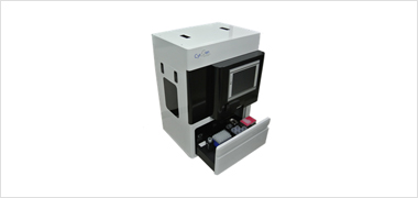
Cell Isolator CIS030
-
Smart Biopsy™ Cell Isolator - CIS 030
Smart Biopsy™ Cell Isolator enriches intact rare cells from human blood and/or body fluid using HDM chip.
(High density microporous chip) - - By size-based filtration, it captures viable cells, which can be useful for downstream application including genomic analysis, immunofluorescent staining, and culture.
- - It is applicable for all kind of body fluid containing rare cells, e.g., blood, pleural effusion, etc.
- - Typical example of enriched rare cells is CTC (circulating tumor cells).
Feature
-
Capture Intact and Pure Cells
Enrich viable cells with high purity by size-based filtration.
-
Automatically detects liquid layer
Detect the volume of the liquid layer by camera and process automatically.
-
Utilize Validated CTC Analysis Workflows
Analyze enriched cells with the downstream assay methods including genomic analysis and immunofluorescent staining.
-
Unique Barcode System
Detect barcode on sample container to avoid intermixing of samples.
-
Variable Volume Sample Loading
Process sample loading in 5~10ml
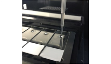
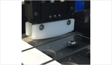
In equipment Workflow
-
STEP 1
Double Negative Selection - Removal of most of erythrocytes and leukocytes in the whole blood
-
STEP 2
HDM Chip - Further removal of remaining smaller nucleated cells by size-based gravity flow filtration
-
STEP 3
CTC Recovery - Retrieval of CTCs
Applications
-
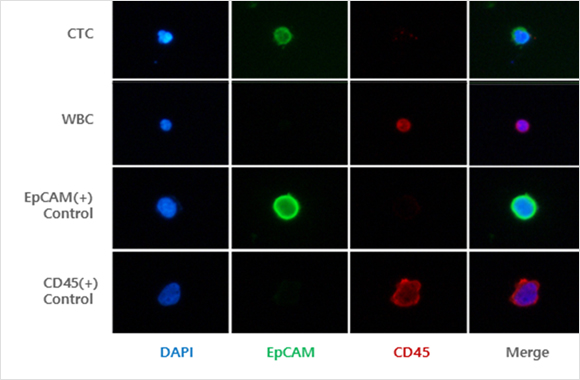
Immunofluorescence Staining
Images of captured CTCs from patients. CTCs were identified by the following
criteria : DAPI (+)/EpCAM (+)/ CD45 (-) -
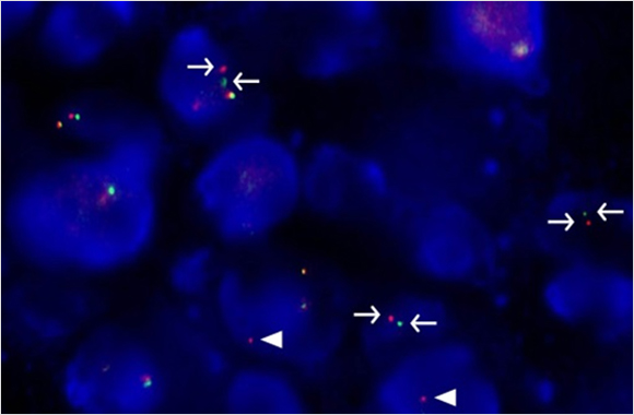
FISH
FISH analysis for the detection of ALK rearrangement
in CTCs from lung cancer patient. Separate red and green signals (arrows)
and isolated red signals (arrow heads) indicates ALK rearrangement. -
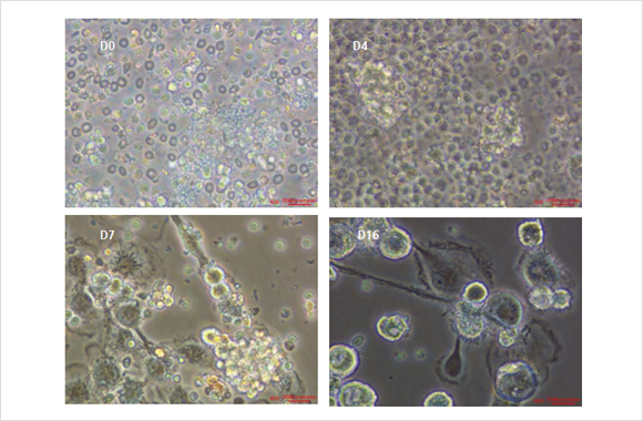
CTC Culture
Representative images of cultured CTCs at day 0, 4, 7 and 16.
-
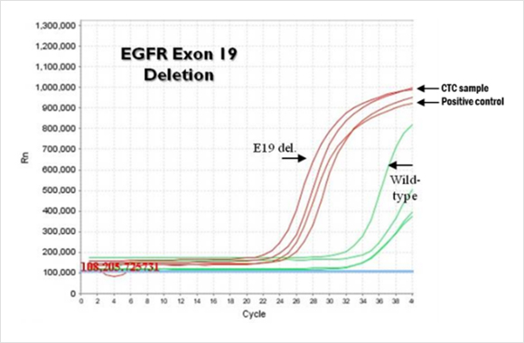
Mutation Analysis in CTC
EGFR exon 19 deletion test with CTCs from lung cancer patient.
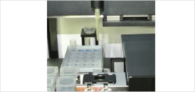
IF Stainer CST030
-
SmartBiopsy™ IF Stainer - CST 030
SmartBiopsy™ IF Stainer is fully automated immunofluorescent staining system, which can stain cells
on the slide with , CD45, anti-EpCAM, CK etc. - - The staining procedure is done in a dark chamber with temperature and moisture control. The system has refrigerated chamber, where reagents are kept.
- - Various staining procedures can be done with various antibodies, and up to 12 slides can processed at one time.
Feature & Performance
-
Accurate volume of reagents
Can release accurate volume of reagents at each step of procedure, and remove reagents on the slides.
-
Automatic mounting
Mount coverslip automatically with DAPI-containing mounting medium.
-
Cold Storage
Refrigerated chamber can maintain the temperature of reagents during the procedure.
-
Temperature control chamber
Automatic temperature & humidity control. External light blocking device.
-
Multiple Sample Loading
Can process 1~12 slides.
-
Covering Module
Automatic cover glass attachment device on the stained sample slide.
-
Applications
Cell staining-based High Throughput screening Development of Staining Protocol.
In equipment Workflow
-
STEP 1
Liquid Vision
-
STEP 2
Liquid Dispensing & Incubation
-
STEP 3
Sample Sealing on Cover glass
Immunofluorescent staining - CIKF10
Immunofluorescent staining image of circulating tumor cells detected from clinical samples.
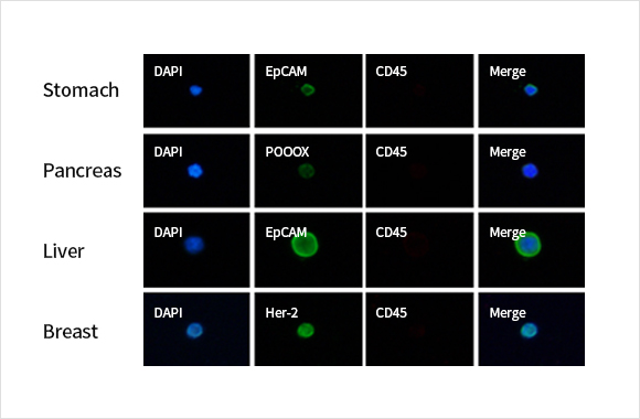
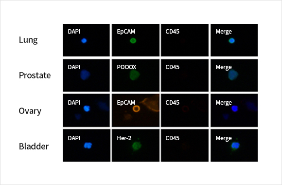
A
Immunofluorescent staining for circulating tumor cell specific biomarkers and leukocyte expression proteins was
performed to identify circulating cancer cells isolated from various cancers.
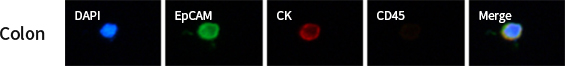
B
4-channel analysis using two different cancer cell-specific biomarkers (EpCAM & CK) is possible using circulating
tumor cells from patients with colorectal cancer.
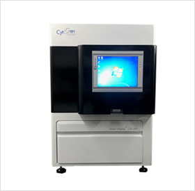
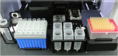
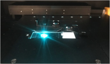
Cell Image Analyzer CIA040
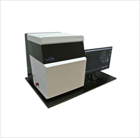
-
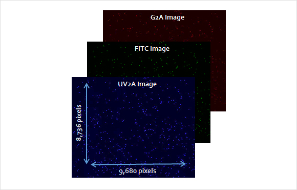
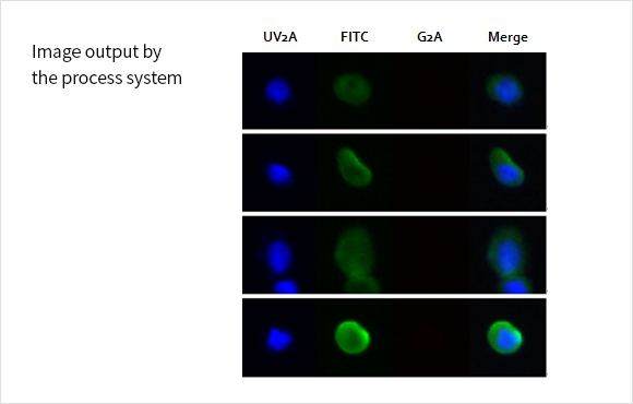
Smart Biopsy™ Cell Image Analyzer - CIA 040
The Smart Biopsy™ Cell Image Analyzer captures immunofluorescent images of cells stained with
anti-EpCAM, -CD45, -CK antibodies. The system includes Image Analyzing software. - - Cell image capture on accurate location provides high-quality merged image of multi-fluorescence.
- - The software provides individual values of intensity and morphology of each cell. Entire slide image and/or magnified image can be obtained.
- - All modules can be operated by both automatic and manual control.
Feature & Performance
-
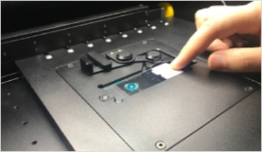
Single Slide Loading
Single slide loading with automatic cover for opening and closing
-
Multiple Color Shot on Same Position
High-quality of merged image.
-
Automatic Analysis
Automatic Sample Image Extraction.
-
Easy-Operating Software
Individual value of fluorescent intensity, cell morphology of each cell. Real time zoom in/out of images on the slide.
Feature & Performance
-
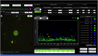
Fluorescence Intensity Analysis
- Analysis of target protein analysis based on immunofluorescence staining
- Analysis of cell morphology
-
Automatic Target Counting
- Automatic counting capacity : more than ten thousands of cells
- Automation : From analysis to Data report
-
Cancer Cell Counting
- Filter mode : For detection of specific cancer cells.
-
In equipment Workflow
-
STEP 1
Sample Loading on platform
-
STEP 2
Focusing & Shooting
-
STEP 3
Analyzing & Reporting